
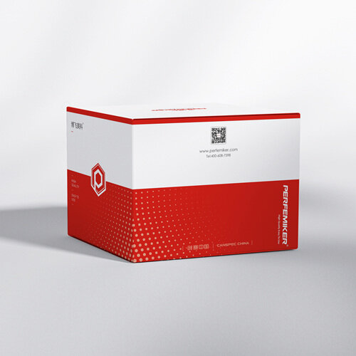
Hematoxylin and Eosin (H&E) Staining Solution
Hematoxylin and Eosin (H&E) staining is the most fundamental and widely used histological technique for tissue sectioning in pathology.
PM20912
Perfumer
100 mL
China
In Stock
On Sale
Share to
Primarily used for staining cell tissues.
Hematoxylin and Eosin (H&E) staining is the most fundamental and widely used technique in histopathology for coloring cell tissues. As a standard method in pathology slide preparation, it is indispensable across biology, histology, pathology, and cytology, playing a critical role in diagnosis, teaching, and research. Cell nuclei, composed of acidic substances, bind strongly to basic dyes like hematoxylin, while the cytoplasm contains more basic components that attract acidic dyes like eosin. Consequently, after H&E staining, cell nuclei appear distinct blue-purple, cytoplasm, muscle fibers, and collagen fibers take on varying shades of red, and red blood cells stain orange-red.
Related Downloads
No download materials available
Recommended Related Products
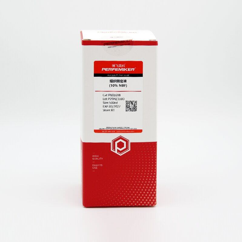
Fixative solution (10% NBF)
Organic fixative (10% NBF), also known as neutral buffered formalin (10%), is composed primarily of formaldehyde, phosphate buffer, and deionized water. It has a pH range of 7.2–7.4 and is suitable for fixing most tissue types.
Hematoxylin and Eosin (H&E) Staining Kit
Hematoxylin and Eosin (H&E) staining colors the cytoplasm red, nuclei blue-purple, and red blood cells orange-red, while other components appear in varying shades of red. It is widely used for staining tissue sections, blood smears, bone marrow sections, and cell smears in immunohistochemistry. As one of the most routine staining methods, H&E is essential in cell biology, histology, and pathology.
Need product inquiries or a discount quote?
Our expert team will provide you with detailed product information and customized solutions.